Presented by: Andrei Iagaru, MD
Covered by: Ivy Riano, M.D., Research Chief Fellow, Hematology and Oncology Unit, Dartmouth Cancer Center, Lebanon, NH.

Professor of Radiology (Nuclear Medicine), Stanford Health Care
Dr. Andrei Iagaru, chief of the Division of Nuclear Medicine and Molecular Imaging at Stanford University Medical Center, discusses the progress of the molecular imaging as an emerging field that integrates advanced imaging technology with cellular and molecular biology. Particularly, the approval and implementation of prostate-specific membrane antigen PET (PSMA-PET) scans has highlighted the advancement of molecular imaging for patients with prostate cancer. PSMA is a transmembrane protein overexpressed in prostate cancer with known enzymatic activities that acts as glutamate-preferring carboxypeptidase [1].
Dr. Iagaru explains that the first PSMA radiopharmaceutical, Gallium 68 PSMA-11 (68Ga-PSMA-11), was FDA approved for PET imaging of PSMA-positive lesions in patients with prostate cancer [2]. Later, the PSMA-PET imaging agent piflufolastat F 18 (18F-DCFPyL; Pylarify) was approved in the same population based on data from the phase 3 CONDOR study [3]. Importantly, the combination of diagnostics and therapy, also known as theragnostics, results in significant survival benefits for patients with prostate cancer who underwent multiple lines of therapy [4].
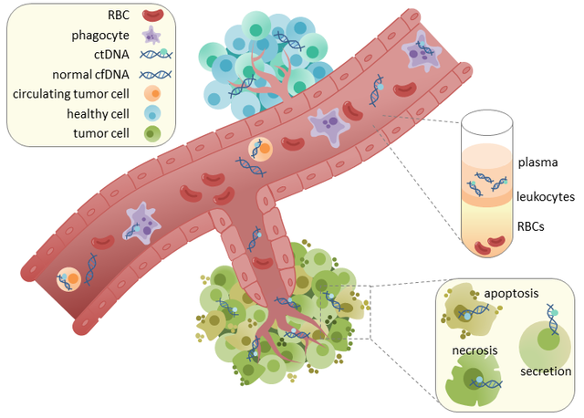
The combination of molecular imaging with omics technologies and analyses of circulating tumor cells is necessary to gain a better understanding of all tumor biology to allow the development of novel therapeutic strategies [5].
- O’Driscoll et al. Pharmacol.2016.
- Park et al. Radiology 2018.
- Morris et al. Clin Cancer Res. 2021.
- Sathekge et al. J Nucl Med 2022.
- Weber et al. JNM 2020.


